Upper Limb Electrical Stimulation After Stroke: Complete Clinician & Patient Guide
NeuroRehab Team
Tuesday, January 6th, 2026
Electrical stimulation for stroke rehabilitation shows promising results for 85% of stroke survivors with arm and hand impairments. Upper limb recovery remains one of the most challenging parts of rehabilitation, despite advances in stroke care. Stroke patients don’t deal very well with simple daily activities like eating, dressing, and personal care.
Clinicians now have powerful tools through electrical stimulation (ES) techniques, especially functional electrical stimulation, to tackle these challenges. The therapy activates weakened or paralyzed muscles by applying controlled electrical impulses, so these muscles work in purposeful patterns. Research proves that ES can substantially improve motor function, reduce pain, and prevent complications like shoulder subluxation when combined with detailed rehabilitation programs.
This piece really gets into how electrical stimulation works for upper limb recovery, available types, and evidence-based protocols to implement it. Readers will learn about proper device setup, safety considerations, and patient education strategies. Both clinicians and patients will get a full picture of how to make this therapy work in stroke recovery programs.
Understanding Electrical Stimulation After Stroke
Electrical stimulation offers a groundbreaking way to help stroke survivors regain their upper limb function. About 40% of stroke survivors can’t use their upper limbs properly, which leads to disability and affects their quality of life [1]. Let’s explore how electrical stimulation therapy works, why it matters for upper limb recovery, and its role in neuroplasticity.
What is electrical stimulation?
Electrical stimulation (ES) helps rehabilitate patients by using controlled electrical impulses that activate specific muscles and nerves. The process is simple – doctors place non-invasive electrodes on the skin above targeted muscle groups [2]. These electrodes deliver carefully adjusted electrical impulses to the tissues underneath, which makes muscles contract in a controlled way [3].
ES works as a substitute for your brain’s natural electrical signals that usually trigger movement. After a stroke, damaged areas of the brain can’t send these signals to muscles effectively, which results in weakness or paralysis [2]. The electrical impulses from ES create a temporary bridge across this communication gap. This allows muscle activation even when patients have limited voluntary control.
Doctors can adjust the strength and pattern of stimulation based on each patient’s needs and comfort levels. The stimulation ranges from gentle muscle contractions that provide sensory feedback to stronger movements that help with joint motion and everyday activities [3].
Why upper limb recovery matters
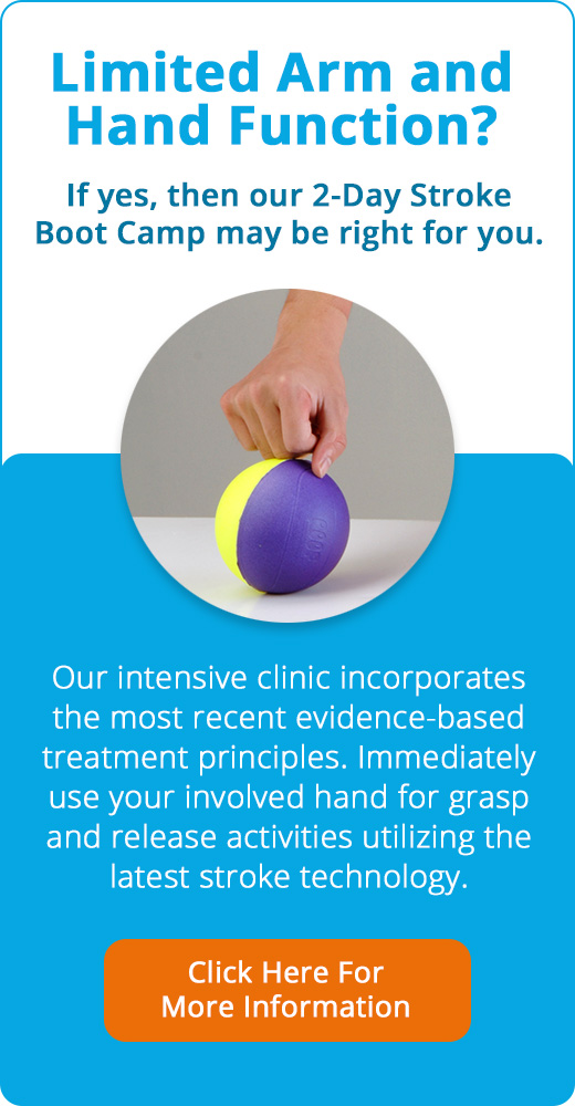
Upper extremity rehabilitation after stroke needs special attention for good reasons. The biggest concern is that 40% of stroke survivors end up with a hand they can’t use, which makes everyday tasks difficult [1]. This becomes especially challenging because most personal care activities need both hands to work together.
Problems with upper limbs affect independence more than lower limb issues, which mainly impact walking. Research shows that while early rehabilitation focuses on walking, many stroke survivors later realize how important arm and hand function is to stay independent [1]. Studies indicate that after twelve months, survivors become more worried about their upper limb function as they understand its basic importance for self-care and meaningful activities [1].
Recent systematic reviews looked at data from 2,774 subacute stroke patients and found encouraging results. Upper extremity function showed clear improvements over time, with Fugl-Meyer Assessment of Upper Extremity (FMA-UE) scores improving by an average of 10 points at 4 weeks and up to 16 points at 24 weeks from where they started [4]. These findings show both the potential for recovery and why we need effective upper limb rehabilitation strategies.
How ES supports neuroplasticity
ES taps into the brain’s natural ability to reorganize itself—a process called neuroplasticity. When muscles receive electrical stimulation, they send sensory feedback to the brain. This activates damaged neural pathways and helps form new connections [2].
The process works through several key mechanisms:
- Neural activation: ES sends input to affected neural pathways and helps activate parts of the brain damaged by stroke [2].
- Synaptic strengthening: Regular stimulation makes existing synaptic connections stronger and creates new ones. Research shows that ES combined with physical exercise can boost motor cortex synaptic connectivity up to 200% more than exercise alone [5].
- Specific plasticity induction: Using ES alongside intentional movement or training creates specific plasticity in the corticospinal network—the vital pathway for controlling voluntary movement [5].
The principle “neurons that fire together, wire together” plays a key role here. Stroke patients who try to move while receiving ES create synchronized activation that boosts neuroplastic changes [6]. This explains why active participation during ES therapy works better than passive stimulation.
ES also increases neurotrophins and boosts the expression of neuronal proteins like MAP2, which help dendritic growth and neural reorganization [7]. These biological changes help restore motor function by changing structures in the brain and spinal cord.
Types of Electrical Stimulation Used in Stroke Rehab
Different electrical stimulation methods have evolved to help with upper extremity rehabilitation after stroke. Each type gives unique benefits and works differently. Clinicians can choose the best method based on what their patients need and their recovery goals.
Functional Electrical Stimulation (FES)
FES applies electrical pulses to motor neurons. This causes muscle contraction that creates or increases movement around a joint [8]. FES stands out because it includes voluntary movement, which makes it different from passive stimulation methods [8]. This active participation is vital because neuroimaging studies showed greater blood flow to the ipsilesional sensory-motor cortex when patients used volitional FES compared to passive electrical stimulation [8].
We can classify FES systems by how patients give feedback. Therapists manually control open-loop FES systems with preset patterns. Closed-loop FES systems use brain-computer interfaces (BCI) or electromyography (EMG) for up-to-the-minute control [9]. Research showed good results from FES-based rehabilitation. Patients improved their Fugl-Meyer Assessment scores (manually controlled FES: mean difference = 5.6, 95% CI [3.77, 7.5], P < 0.001; BCI-controlled FES: mean difference = 5.37, 95% CI [4.2, 6.6], P < 0.001; EMG-controlled FES: mean difference = 14.14, 95% CI [11.72, 16.6], P < 0.001) [9].
Neuromuscular Electrical Stimulation (NMES)
NMES uses short electrical pulses through surface electrodes to excite peripheral nerves and make muscles contract [5]. NMES typically uses pulse frequencies of 10-100 Hz, amplitudes of 10-120 mA, and pulse widths of 200 μs to 1 ms [5]. Higher frequencies create larger forces but quickly tire the muscles [5].
Cyclic NMES (cNMES) is a common method. The stimulation cycles on and off repeatedly, and clinicians set the timing, repetitions, and intensity [8]. cNMES is easy to give but doesn’t need the patient to participate actively [8]. Notwithstanding that, it helps improve upper limb motor scores and strength [10]. One study showed that just 10 hours of NMES with regular rehabilitation improved arm function in acute stroke patients by a lot [10].
Transcutaneous Electrical Nerve Stimulation (TENS)
Doctors have used TENS in stroke rehabilitation for decades. It started as a pain management tool but now helps with motor recovery [3]. TENS over the paretic limb reduces stretch reflex magnitude and extends H-reflex latency at the peripheral level [3]. The brain areas that match stimulated body parts become excited at the cortical level, which creates greater activation of the lesioned hemisphere [3].
Unilateral TENS (Uni-TENS) with task-oriented training works better than placebo stimulation. It increases wrist flexor strength, reduces spasticity, and helps motor control [3]. Bilateral TENS (Bi-TENS) on both paretic and non-paretic limbs provides extra sensory input. This helps balance interhemispheric inhibition and activates neural networks in both hemispheres [3]. Recent studies confirm that TENS sessions longer than 30 minutes help improve spasticity in stroke patients [3].
Transcutaneous Electrical Acupoint Stimulation (TEAS)
TEAS is a new non-invasive therapy that combines transcutaneous electrical stimulation with traditional acupuncture principles [11]. It uses low-frequency pulsed direct current to stimulate peripheral acupoints and surrounding tissues. This helps information transmission to the central nervous system and improves local neuromuscular function [11].
TEAS doesn’t cause discomfort like pain and bleeding, unlike electroacupuncture or traditional acupuncture. This leads to better patient compliance [11]. Clinical practice shows that TEAS with rehabilitation training improves muscle strength, hand function, grip strength, and manual dexterity while easing pain and muscle spasms [11]. A newer network meta-analysis found that TEAS had the best effect on upper limb motor recovery after stroke [11].
EMG-triggered and reciprocal stimulation
EMG-triggered electrical stimulation is an advanced method that connects muscle stimulation to the patient’s own movement attempts [4]. Surface EMG recording electrodes on target muscles detect signals when patients try to move [4]. The electrical stimulation starts once the processed EMG signal goes above a preset threshold [4].
This method is nowhere near passive stimulation because it needs active participation. Many researchers believe this improves neuroplasticity and motor relearning [1]. Studies show EMG-triggered FES helps improve Box and Block test scores, finger-extension strength, and movement quality [1]. There’s another reason to be excited – bilateral protocols that combine unimpaired limb movements with active stimulation on the impaired limb show promising results [1].
Contralaterally controlled functional electrical stimulation (CCFES) brings a new approach. The patient’s volitional opening of their unaffected hand controls stimulation intensity to the paretic hand [8]. Patients can control when and how much their hand opens during tasks. This shows better results in upper extremity hemiplegia compared to regular NMES [12].
How Electrical Stimulation Works in the Body
The way electrical stimulation works to restore movement in stroke-affected limbs depends on specific physiological mechanisms. Medical professionals need to learn about these mechanisms to achieve the best rehabilitation results.
Muscle contraction and nerve activation
Our nervous system uses electrical signals as a natural way to control muscles. Neurons send information through action potentials—brief electrical impulses that show changes in cell potential of approximately 80-90 mV [13]. Brain areas damaged by stroke can’t generate these signals correctly, which results in weakness or paralysis [7].
Electrical stimulation helps by creating artificial action potentials in peripheral nerves. The electrical current starts by causing a localized depolarization of the nerve cell membrane. This triggers an action potential that travels in two directions at once [13]:
- Orthodromic propagation: Action potentials travel toward the muscle, which causes contraction
- Antidromic propagation: Action potentials travel toward the central nervous system
These electrical impulses reach the neuromuscular junction and release acetylcholine. This chemical binds to receptors on the muscle fiber and makes the muscle contract. The process copies the natural electrical commands that would normally come from the brain [7].
Motor unit recruitment and fatigue
The main difference between electrically-stimulated and voluntary muscle contractions shows up in how motor units get recruited. This explains both the advantages and limits of electrical stimulation in stroke rehabilitation.
Voluntary contractions follow a specific order in the nervous system:
- Small-diameter, slow-twitch (Type I) fibers activate first
- Medium-diameter fibers follow
- Large-diameter, fast-twitch (Type II) fibers activate last [14]
Electrical stimulation works the opposite way. Research shows that ES recruits motor units in a “nonselective, spatially fixed, and temporally synchronous pattern” [15]. The largest-diameter fibers activate first because they have lower electrical thresholds. These fibers create quick, powerful contractions [14].
This reversed order explains why muscles get tired faster with electrical stimulation than with voluntary movement. The fast-twitch fibers that activate first tire quickly, so therapists must carefully adjust stimulation settings to reduce fatigue [15].
The frequency of stimulation plays a big role in how quickly muscles tire. Frequencies above 50 Hz create stronger muscle forces but lead to faster fatigue and reduced contraction force [16]. The pulse width also matters—wider pulses create stronger responses in both the brain and muscles [16].
Surface vs percutaneous electrodes
The choice of how to deliver electrical stimulation affects both treatment results and patient comfort. Therapists can pick from several electrode types based on their goals and what patients need.
Surface electrodes are the most common choice. These non-invasive devices stick to the skin above target nerves or muscles [13]. They’re easy to apply, simple to adjust, and cost less than other options. The current needed for surface electrodes ranges from 2-120 mA to get through skin resistance [2].
Percutaneous electrodes offer a way to stimulate deeper tissues more precisely. These thin wires go through the skin into muscle tissue near target nerves [13]. They need less current—usually around 25 mA—and offer more precise stimulation [2]. However, these electrodes might cause infections and need special care.
Fully implanted electrodes give the most permanent option. Doctors surgically place them near specific nerves for precise, long-term stimulation [13]. Though surgery is required, these systems target muscles most accurately with minimal current spread to nearby tissues.
Surface electrodes work best for most upper extremity stroke rehabilitation. They’re safe, effective, and practical. Their non-invasive nature helps patients start treatment early, which often helps them recover better [2].
Clinical Applications for Upper Limb Recovery
Electrical stimulation has emerged as a great way to help stroke survivors. It addresses upper limb problems that affect their independence and quality of life. Research shows it works well in several key areas of rehabilitation.
Reducing shoulder subluxation
Shoulder subluxation affects up to 81% of individuals after stroke. This condition often leads to pain and poor upper limb function [17]. Clinical evidence supports the use of electrical stimulation right after stroke to prevent rather than reduce existing subluxation. Adding ES to regular therapy prevents shoulder subluxation by an average of 6.5mm [17].
A study of 40 patients who received stimulation within 48 hours after their stroke showed much less subluxation after 4 weeks of treatment [18]. The best results come from stimulating the supraspinatus and posterior deltoid muscles. These muscles play a vital role in keeping the glenohumeral joint properly aligned [19]. Studies using electromyography have identified these muscles as the main stabilizers of the shoulder joint [19].
Improving motor control and coordination
Upper extremity electrical stimulation boosts motor recovery through different feedback systems. EMG-triggered stimulation works better than passive stimulation alone. This happens because patients start the movement before getting assistance [20].
Research reveals notable improvements in functional measures:
- FMA scores improved with manually controlled FES (mean difference = 5.6) [21]
- Brain-computer interface controlled FES showed similar benefits (mean difference = 5.37) [21]
- EMG-controlled FES produced the best results (mean difference = 14.14) [21]
Bilateral TENS treatment on both affected and unaffected limbs provides extra sensory input. This helps balance interhemispheric inhibition and works better than single-side applications [6].
Enhancing range of motion and strength
Electrical stimulation helps improve range of motion in stroke-affected limbs substantially. Research shows that NMES combined with other treatments increased ROM by an average of 2.87 degrees [5]. The benefits vary by location. Leg stimulation increased ROM by 3.13 degrees, and elbow applications showed a 4.57-degree improvement. However, wrist applications had little effect [5].
These improvements take time to develop. Studies show that patients need at least 10 hours of NMES along with standard rehabilitation [22]. This treatment strengthens weak muscles and improves voluntary control and function.
Managing spasticity and pain
One in five stroke patients develops spasticity. This condition limits mobility and makes it hard to use affected limbs, affecting quality of life [23]. Neuromuscular electrical stimulation helps reduce this problem. Studies show that NMES, used alone or with other treatments, reduces spasticity scores by -0.30 on the Modified Ashworth Scale [5].
Different stimulation settings work well, with frequencies between 18-50 Hz and pulse durations from 0.1-0.4 ms [5]. Transcutaneous Electrical Acupoint Stimulation (TEAS) shows excellent results in reducing spasticity [24]. The treatment also helps with pain management, as many studies report lower pain levels after electrical stimulation [23].
Device Setup and Treatment Parameters
Getting the best results from electrical stimulation in stroke recovery depends on setting up the device correctly and choosing the right parameters. Medical practitioners need to set these elements carefully to get the best therapeutic results and keep patients comfortable and safe.
Electrode placement and configuration
The right electrode placement makes a big difference in how well the stimulation works. To help with wrist extension, put one electrode on the dorsal forearm, 2-3 finger-widths below the lateral epicondyle, and place the second electrode over the extensor tendons near the wrist [9]. Shoulder subluxation treatment needs electrodes placed over the supraspinatus and posterior deltoid muscles [8]. Elbow extension works best with one electrode on the mid-triceps and another near the distal triceps tendon just above the elbow [9]. The skin surface must be clean and dry before you start – use alcohol prep pads if needed [25].
Cyclical vs EMG-triggered modes
You can deliver stimulation through cyclical or EMG-triggered methods. Cyclical stimulation uses preset on-off patterns, usually starting with 10 seconds on and 10 seconds off. As patients build tolerance, this can move to 10 seconds on and 5 seconds off [8]. EMG-triggered stimulation works differently – it activates only when patients try to move beyond a preset threshold [21]. Research shows EMG-triggered stimulation leads to slightly better results than cyclic stimulation (4.2 vs. 2.3 points on the Action Research Arm test), though researchers didn’t find this statistically significant [26].
Adjusting frequency, pulse width, and intensity
The right parameter settings play a crucial role in treatment success:
- Frequency: Upper limb applications usually need 18-50 Hz [5]. Lower frequencies (1-10 Hz) create mild muscle twitches, while higher ones (60-70 Hz) give stronger contractions but tire muscles faster [25].
- Pulse width: Most settings fall between 0.1-0.4 ms [5]. Wider pulses activate more motor neurons but might feel uncomfortable [25].
- Intensity: Start with low settings and slowly increase until you see the limb move, making sure the patient stays comfortable [8].
Treatment duration and dosage guidelines
Most effective treatments last 20-60 minutes per session [4]. Clinical trials typically run 45-60 minute sessions 3-5 days each week for 8-16 weeks, adding up to about 40 sessions [10]. Just 10 hours of total stimulation combined with regular rehab can improve function substantially [27]. A notable study revealed that motor outcomes in subacute stroke didn’t depend on how long EMG-triggered stimulation lasted, so doctors can adjust treatment time based on what patients can handle [28].
Safety, Contraindications, and Patient Education
Electrical stimulation provides remarkable benefits for stroke recovery, but safety must come first in clinical practice. Clinicians need a full picture of key contraindications to deliver treatments that work without risking patient safety.
Who should not use ES
Patients with cardiac pacemakers or implantable cardioverter defibrillators should never receive upper extremity electrical stimulation because it might cause device malfunction [29]. The treatment should be avoided over:
- Malignancy sites (tumor growth might accelerate)
- The gravid uterus during pregnancy, especially in the first trimester
- Active deep vein thrombosis
- Open wounds, tumors, or the anterior neck region
Patients with impaired sensation, active infection, or cognitive deficits that limit their ability to give feedback should also be carefully evaluated [29].
Common side effects and how to manage them
Functional electrical stimulation usually causes minor adverse events. Patients commonly report tingling (37.3% of papers), burning sensation (18.7%), headaches (14.7%), and fatigue (14.7%) [30]. The skin might become red where electrodes are placed, but this typically goes away within an hour [3].
You can try hypoallergenic electrodes or adjust current levels if skin irritation occurs [3]. Keep in mind that stimulation should never hurt—it should just feel uncomfortable [31].
Educating patients and caregivers
The first step in patient instruction is explaining what they should expect to feel. Tell them directly: “You should feel tingling or muscle contractions, but report anything else immediately” [32]. The electrodes must make full contact with the skin to prevent current concentration [25].
Consent and documentation best practices
Using standardized forms helps identify all possible contraindications [29]. Documentation should include informed consent, clinical reasoning behind treatment decisions, protocol adjustments, and how patients respond to treatment [29].
Conclusion
Electrical stimulation therapy is a powerful tool that helps stroke survivors overcome upper limb challenges. Research shows it works well when integrated into complete stroke recovery programs. Clinicians can choose from several ES modalities to meet specific patient needs. These include functional electrical stimulation, neuromuscular electrical stimulation, and transcutaneous electrical nerve stimulation.
All the same, this therapy needs careful attention to electrode placement, stimulation parameters, and treatment protocols. The core team must choose the right frequencies, pulse widths, and intensities based on each patient’s condition and goals. On top of that, it will give a safe and effective treatment when clinicians spot contraindications and manage potential risks.
The therapy helps improve neuroplasticity – the brain’s remarkable way to reorganize and create new neural connections. This rewiring of the brain leads to meaningful recovery, especially when patients actively participate in their therapy. Combining voluntary movement with electrical stimulation gives better results than passive approaches.
Stroke patients who don’t deal very well with simple self-care tasks can see big improvements. These include better motor control, muscle strength, range of motion, and coordination through proper ES therapy. While results differ from person to person, evidence shows electrical stimulation is valuable for upper limb rehabilitation. Then, both clinicians and patients should think over this option as part of their complete recovery plan.
Without doubt, electrical stimulation is just one piece of the rehabilitation puzzle. Yet, knowing how to target key problems – from shoulder subluxation to spasticity – makes it essential to maximize upper limb recovery after stroke. Early treatment, consistent use, and proper adjustment of parameters help stroke survivors regain independence and boost their quality of life through improved upper extremity function.
Key Takeaways
Electrical stimulation offers stroke survivors a scientifically-backed pathway to regain upper limb function, with 85% experiencing arm impairments that significantly impact daily independence.
• Multiple ES types target different recovery needs: FES for functional movement, NMES for muscle strengthening, TENS for pain/spasticity management, and EMG-triggered systems for active participation.
• Active patient participation amplifies results: EMG-triggered stimulation produces 14.14-point improvements in motor scores versus 5.6 points with passive stimulation alone.
• Proper setup determines success: Electrode placement over specific muscle groups, frequencies of 18-50 Hz, and 20-60 minute sessions 3-5 times weekly optimize therapeutic outcomes.
• Early intervention prevents complications: ES applied within 48 hours post-stroke prevents 6.5mm of shoulder subluxation and supports crucial neuroplasticity during recovery windows.
• Safety screening is essential: Absolute contraindications include pacemakers and pregnancy, while proper parameter adjustment prevents skin irritation and ensures patient comfort throughout treatment.
When combined with conventional rehabilitation, electrical stimulation harnesses the brain’s neuroplasticity to rebuild neural pathways damaged by stroke. The key lies in selecting appropriate stimulation types, maintaining consistent treatment schedules, and ensuring active patient engagement to maximize functional recovery outcomes.
References
[1] – https://www.jptrs.org/view.html?volume=2&number=1&spage=1
[2] – https://pmc.ncbi.nlm.nih.gov/articles/PMC8984173/
[3] – https://strokengine.ca/en/interventions/functional-electrical-stimulation-lower-extremity/
[4] – https://www.neurorehabdirectory.com/blog-stroke-recovery-electrical-stimulation-electrode-placement/
[5] – https://www.ahajournals.org/doi/10.1161/strokeaha.115.009633
[6] – https://www.ahajournals.org/doi/10.1161/STROKEAHA.121.036895
[7] – https://www.flintrehab.com/electrical-stimulation-for-stroke-patients/?srsltid=AfmBOop03dmrk0QxTkFkO6P9f8JFu5aDFo_CqiAxCYDMexfs_WluaqBv
[8] – https://www.medicaljournals.se/jrm/content/html/10.2340/16501977-0917
[9] – https://www.valkyrie-vr.com/blog/fes-electrode-placements-for-upper-limb-stroke-recovery1
[10] – https://www.frontiersin.org/journals/neuroscience/articles/10.3389/fnins.2020.00718/full
[11] – https://www.frontiersin.org/journals/aging-neuroscience/articles/10.3389/fnagi.2024.1438994/full
[12] – https://pubmed.ncbi.nlm.nih.gov/35574940/
[13] – https://en.wikipedia.org/wiki/Functional_electrical_stimulation
[14] – https://www.physio-pedia.com/Neuromuscular_and_Muscular_Electrical_Stimulation_(NMES)
[15] – https://pubmed.ncbi.nlm.nih.gov/21870119/
[16] – https://pmc.ncbi.nlm.nih.gov/articles/PMC7005350/
[17] – https://www.sciencedirect.com/science/article/pii/S0004951414601653
[18] – https://pubmed.ncbi.nlm.nih.gov/10229728/
[19] – https://www.ahajournals.org/doi/10.1161/01.str.30.5.963
[20] – https://pmc.ncbi.nlm.nih.gov/articles/PMC5048487/
[21] – https://www.frontiersin.org/journals/neurology/articles/10.3389/fneur.2023.1272992/full
[22] – https://pmc.ncbi.nlm.nih.gov/articles/PMC6465149/
[23] – https://pmc.ncbi.nlm.nih.gov/articles/PMC9800032/
[24] – https://pmc.ncbi.nlm.nih.gov/articles/PMC8450164/
[25] – https://www.occupationaltherapy.com/articles/stroke-electrical-stimulation-and-therapy-5700
[26] – https://pubmed.ncbi.nlm.nih.gov/18678569/
[27] – https://www.uhd.nhs.uk/uploads/services/docs/stroke/hi-reps/RCP_Guidelines_Estim.pdf
[28] – https://www.tandfonline.com/doi/abs/10.1080/09638288.2025.2535468
[29] – https://www.physio-pedia.com/Electrotherapy_Contraindications
[30] – https://pubmed.ncbi.nlm.nih.gov/35353649/
[31] – https://www.flintrehab.com/electrical-stimulation-for-stroke-patients/?srsltid=AfmBOoqDszNyXcEA30yDOriFujYApj69LZ4-DcKATVfEbuQSx3DdwMZD
[32] – https://www.canberrahealthservices.act.gov.au/__data/assets/word_doc/0010/1981297/Electrical-Stimulation-as-a-Treatment-Modality-for-Adult-Patients-with-Neurological-Conditions.docx

Leave a Reply
You must be logged in to post a comment.